A Safer Approach for High-Risk Wisdom Tooth Removal
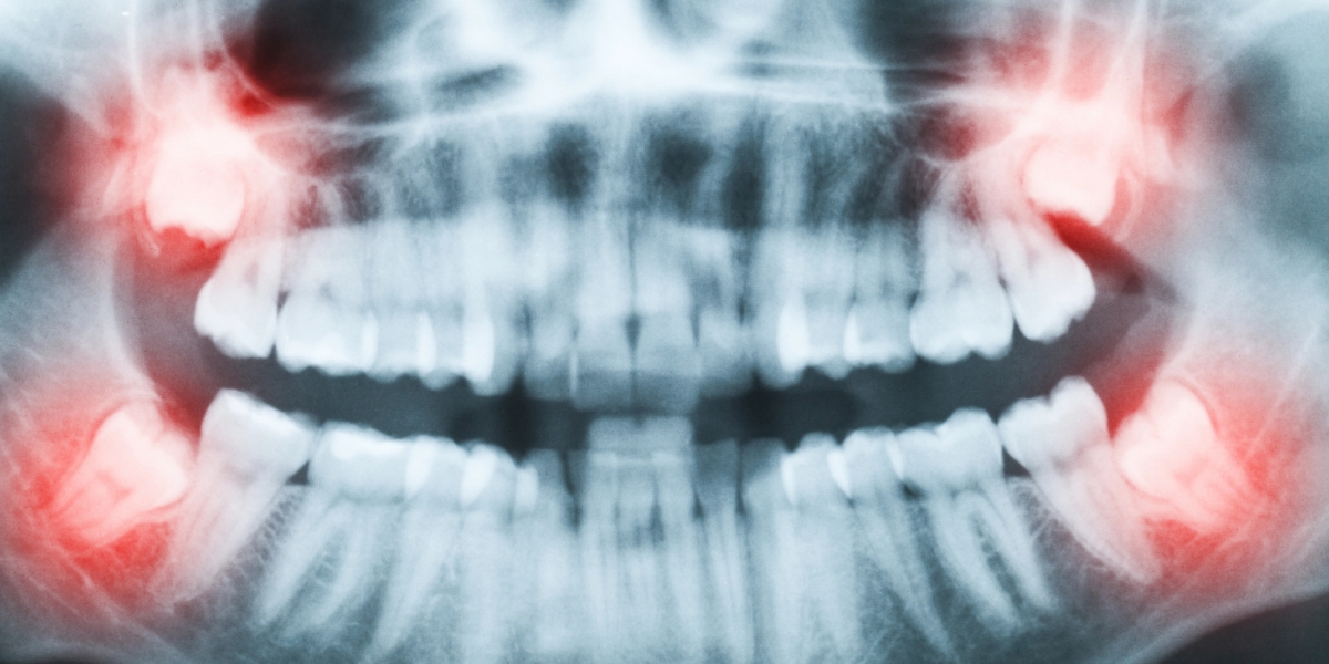
For some patients, removing lower (mandibular) wisdom teeth poses a significant risk to nearby nerves—specifically the inferior alveolar nerve (IAN) and lingual nerve. At Clackamas Implant & Oral Surgery Center, we offer dental coronectomy as an advanced, nerve-sparing alternative for patients with deeply impacted lower third molars.
This highly specialized procedure is performed by Dr. Brett Sullivan, DMD, MD, a dual-degreed oral and maxillofacial surgeon with extensive training and experience. Dr. Sullivan has successfully performed over 1,000 coronectomies, making him one of the most experienced providers in the Pacific Northwest.
What Is a Coronectomy?
A coronectomy involves removing only the crown (top portion) of the impacted wisdom tooth, while leaving the roots in place. This technique is recommended when diagnostic imaging shows that the tooth roots are in close proximity to the IAN or lingual nerve, increasing the risk of nerve damage during full extraction.
By preserving the roots, the procedure allows us to:
- Avoid direct contact with critical nerves
- Prevent common complications like infection, cysts, or pressure on adjacent teeth
- Minimize surgical trauma and support faster recovery
Why Dr. Sullivan May Recommend a Coronectomy
- ✅ Nerve Preservation: The top priority is to protect the inferior alveolar and lingual nerves—especially in cases with high-risk anatomy.
- ✅ Reduced Trauma: Coronectomy is less invasive than full extraction, often resulting in quicker healing and fewer complications.
- ✅ Evidence-Based Decision Making: This treatment is only recommended when advanced imaging—such as cone beam CT (CBCT)—reveals significant anatomical risk.
Dr. Sullivan’s decision to perform a coronectomy is based on radiographic evidence and guided by peer-reviewed clinical standards—not performed arbitrarily.
Understanding the Risk of Nerve Injury
Wisdom tooth removal near nerve structures can carry the following risks:
| Complication Type | Estimated Incidence |
| Temporary IAN Injury | 0.26% – 8.4% |
| Permanent IAN Injury | Up to 3.6% |
| Lingual Nerve Damage | 0.1% – 22% |
These statistics are supported by peer-reviewed studies and systematic reviews, including works by Renton (2010), Guerrero (2014), and Pogrel (2004)—leaders in oral surgery literature.
What to Expect During a Coronectomy
1. Advanced Imaging & Evaluation
Dr. Sullivan will use 3D CBCT scans to assess the exact location of the tooth in relation to your nerves.
2. Surgical Procedure
Under local anesthesia or IV sedation, the crown is carefully sectioned and removed, leaving the root undisturbed and below the gumline.
3. Post-Operative Monitoring
The retained roots are monitored over time and usually become encapsulated in bone, requiring no further treatment.
Is Coronectomy Right for You?
If your diagnostic imaging reveals a high risk of nerve injury from standard extraction, a coronectomy may offer a safer, evidence-based solution. This decision is always made in your best interest after a thorough evaluation.
Schedule Your Consultation Today
To learn whether a coronectomy is right for you, schedule a consultation with Dr. Brett Sullivan at Clackamas Implant & Oral Surgery Center.
Call (503) 652-8080 or request your consultation online to take the next step toward safe, expert care.